البروفيسور سعيد أختر

Job title: Professor

Department: BMS

Email: sakhtar@inaya.edu.sa

Extension: 227


Education:

Ph.D. (1986 – 1990): University of Reading, U.K;

MTech (1981 – 1983): University of Reading, U.K
Area of Expertise

Prior to coming to the Inaya college, I worked in King Saud University for 12 years and in the UK for 18 years at Reading University; Oxford Brooks University; Oxford Research Unit, Open University, and Cardiff University.

Most of my research involves investigating re-modelling of collagen fibrils and proteoglycans in normal and pathological cornea such as keratoconus, post-LASIK cornea, cross-linked cornea, and corneal dystrophies. We are the 1st research group who published the origin of the keratoconus from the peripheral cornea.

Human cornea has five layers. I have discovered a new layer of the peripheral cornea. The layer is called pre-endothelial layer (PENL).

Recently, Dr Sami encouraged high profile research and authorized to purchase of state-of-the-art JEOL transmission electron microscopy technology.  Prof Akhtar is carrying out a ground breaking research on the cornea with help of the transmission electron microscope attached with a 24-megapixel camera. With new technology, I have explained the structure of PENL with 3D tomography on transmission electron microscope (Figure 1, 2).
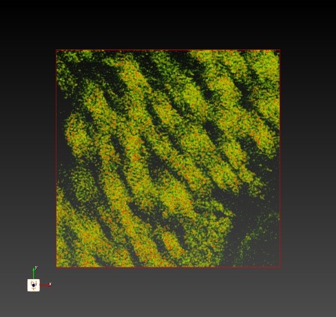
3D image of collage fibrils present in the pre-endothelial layer.
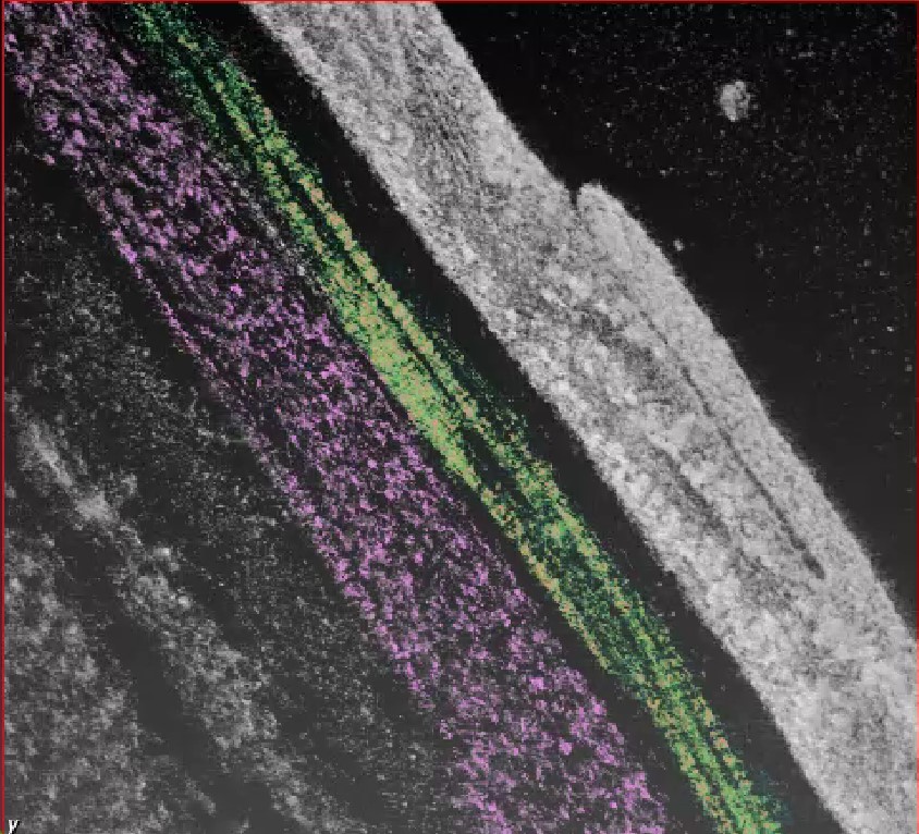
3D image of pre-endothelial layer

I have also investigated the role of collagen fibrils and proteoglycans of intervertebral disc, bicuspid heart valve and arthritis cartilage. Last few years I have been carrying out research on optic nerve and of glaucoma eyes and published in Q1 journal in collaboration with Finland and Poland.

Research and Publications

Relevant Publications in Q1  

  1. Saeed Akhtar, Adrian Smedowski, Adnan Ali Khan, Hind Debasi, Hanan Mofty, Ramachandran Samivel, Turki Almubrad (2024). Glycosaminoglycans and collagen fibril distribution at various depths of the corneal stroma of normal and CXL treated rats. Exp Eye Res. 239:109780. doi: 10.1016/j.exer.2024.109780.

  2. Anna Pacwa, Joanna Machowicz, Saeed Akhtar, Piotr Rodak, Xiaonan Liu1, Marita Pietrucha-Dutczak, Joanna Lewin-Kowalik, Marialaura Amadio6 and Adrian Smedowski (2023). Deficiency of the RNA-binding protein ELAVL1/HuR leads to the failure of endogenous and exogenous neuroprotection of retinal ganglion cells. Frontiers in Cellular Neuroscience 17:1131356. DOI 10.3389/fncel.2023.1131356

  3. Saeed Akhtar, Ramachandran Samivel, Adrian Smedowski, Aljoharah Alkanaan, Ali Masmali, Omar Kirat, Adnan Ali Khan, Turki Almubrad (2022). A unique pre‑endothelial layer at the posterior peripheral cornea: ultrastructural study. Sci Rep15;12(1):2556. doi.org/10.1038/s41598-022-06552-6

  4. Saeed Akhtar, Adrian Smedowski, Ali Masmali, Aljoharah Alkanaan, Adnan Ali Khan, Essam Almutleb, Hanan Khalid Mofty, Hind I Al-Debasi, Ramachandran Samivel, Turki Almubrad (2022). Effect of Ultraviolet-A and Riboflavin treatment on the architecture of the center and periphery of normal rat cornea: 7 days post treatment. Exp Eye Res. 219:109064. doi: 10.1016/j.exer.2022.109064.

  5. Ramachandran Samivel, Umadevi Subramanian, Adnan Ali Khan, Omar Kirat, Ali Masmali, Turki Almubrad, Saeed Akhtar (2022). Lipopolysaccharide Enhances Genotoxicity by Activating GADD45G and NF- κ B in Human Corneal Epithelial Cells. Oxid Med Cell Longev 2022:4328116. DOI: 10.1155/2022/4328116

  6. Ramachandran Samivel, Rajendra Prasad Nagarajan, Umadevi Subramanian,Adnan Ali Khan, Ali Masmali , Turki Almubrad, and Saeed Akhtar (2020). Inhibitory Effect of Ursolic Acid on Ultraviolet B Radiation-Induced Oxidative Stress and Pro-inflammatory Response- Mediated Senescence in Human Skin Dermal Fibroblasts. Oxidative Medicine and Cellular Longevity Volume 2020, Article ID 1246510, 17 pages, https://doi.org/10.1155/2020/1246510.

  7. Smedowski A, Akhtar S, Liu X, Pietrucha-Dutczak M, Podracka L, Toropainen E, Alkanaan A, Ruponen M, Urtti A, Varjosalo M, Kaarniranta K, Lewin-Kowalik J (2020). Electrical synapses interconnecting axons revealed in the optic nerve head – a novel model of gap junctions’ involvement in optic nerve function. Acta Ophthalmol.98:408-417. doi: 10.1111/aos.14272.

  8. Alkanaan A, Barsotti R, Kirat O, Khan A, Almubrad T, Akhtar S (2019). Collagen fibrils and proteoglycans of peripheral and central stroma of the keratoconus cornea – Ultrastructure and 3D transmission electron tomography. Sci. Rep. 2019 Dec 27;9(1):19963. doi: 10.1038/s41598-019-56529-1

  9. Patnaik SR, Zhang X, Biswas L, Akhtar S, Zhou X, Kusuluri DK, Reilly J, May-Simera H, Chalmers S, McCarron JG, Shu X. (2018). RPGR protein complex regulates proteasome activity and mediates store-operated calcium entry. Oncotarget. 9(33):23183-23197

  10. Albalawi A1, Alhasani RHA2, Biswas L3, Reilly J3, Akhtar S4, Shu X5. (2018). Carnosic acid attenuates acrylamide-induced retinal toxicity in zebrafish embryos. Exp Eye Res. 2018 Oct;175:103-114

  11. Smedowski Adrian, Liu Xiaonan, Podracka Lucia, Akhtar Saeed, Trzeciecka Anna, Pietrucha-Dutczak Marita, Lewin-Kowalik Joanna, Jiu Yaming, Lappalainen Pekka, Urtti Arto, Ruponen Marika, Kaarniranta Kai, Varjosalo Markku, Amadio Marialaura (2018).  Increased intraocular pressure alters cellular distribution of HuR protein in retinal ganglion cells – a possible sign of endogenous neuroprotection failure. Biochim Biophys Acta. 2018;1864:296-306.. Impact factor 5.228.

  12. Rakesh Raghupathy, Xinhua Shu, , Xun Zhang, Fei Liu, Reem Alhasani, Saeed Akhtar, Luyuan Pan, Cecilia Moens, Wenchang Li, Mugen Liu, and Breandan Kennedy (2017).  RRGRIP1 is required for rod outer segment development and ciliary protein trafficking in zebrafish. Scientific reports 7(1):16881; Impact factor 4.259

  13. Alkanaan A, Barsotti R, Kirat O, Almubrad T, Khan A, Akhtar S (2017). Ultrastructural study of peripheral and central stroma of keratoconus cornea. BJ Ophthalmology 10.1136

  14. Szatmári-Tóth M, Kristóf E, Veréb Z, Akhtar S, Facskó A, Fésüs L, Kauppinen A, Kaarniranta K, Petrovski G (2016). Clearance of autophagy-associated dying retinal pigment epithelial cells – a possible source for inflammation in age-related macular degeneration. Cell Death Dis. 7(9):e2367. Impact factor 6.5

  15. Koulikovska M, Mehrdad R, Petrovski G, Veréb Z , Akhtar S, Fagerholm P and Lagali N (2015). Enhanced Regeneration of Corneal Tissue via a Bioengineered Collagen Construct implanted by a Non-disruptive Surgical Technique. Tissue Engineering Part A; Tissue Eng Part A. 2015 Mar;21(5-6):1116-30. Impact factor 4.448

  16. Zoltán Veréb, Réka Albert, Szilárd Póliska, Ole Kristoffer Olstad, Saeed Akhtar, Morten C Moe and Goran Petrovsk (2013). Comparison of upstream regulators in human ex vivo cultured cornea limbal epithelial stem cells and differentiated corneal epithelial cells. BMC Genomics 14:900. Impact factor 1.45

  17. Viiri J, Amadio M, Marchesi N, Hyttinen JM, Kivinen N, Sironen R, Rilla K, Akhtar S, Provenzani A, D’Agostino VG, Govoni S, Pascale A, Agostini H, Petrovski G, Salminen A, Kaarniranta K (2013). Autophagy activation clears elavl1/hur-mediated accumulation of sqstm1/p62 during proteasomal inhibition in human retinal pigment epithelial cells. PLoS One. 2013; 8(7): e69563. Impact factor

  18. ALmubrad T and Akhtar S (2011) Ultrastructure features of camel cornea – collagen fibril and proteoglycans. Veterinary Ophthalmology 17:2283-91. ISSN – 1463-5224, Impact Factor 1.125

  19. Akhtar S, Almubrad TM, Bron AJ, Yusif MHM, Benter IF, Akhtar S (2009)  Effect of EGFR in corneal remodelling in diabetes. Acta Ophthalmol 87:881-889. ISSN – 655-665, Impact Factor 2.441

  20. Akhtar S, Kerr BC, Hayes AJ, Hughes CE, Meek KM, Caterson B. (2008) Immunochemical localization of keratan sulfate proteoglycans in cornea, sclera, and limbus using a keratanase-generated neoepitope monoclonal antibody. Invest Ophthalmol Vis Sci. 49:2424-2431. ISSN – 0146-0404, Impact Factor 3.431

  21. Akhtar S, Schonthaler HB, Bron AJ and Dahm R. (2008) The ultrastructural organization of stromal collagen and proteoglycan in the zebrafish cornea during development. Acta Ophthalmol 86:655-665. ISSN – 655-665, Impact Factor 2.441

  22. Akhtar S, Bron AJ , Salvi SM, Hawksworth NR, Tuft SJ and Meek KM. (2008) Collagen fibrils and proteoglycans in keratoconus – a quantitative ultra-structural analysis.  Acta Ophthalmol 86:764-772

  23. Albon J, Farrant S, Akhtar S, Young R, Boulton ME, Smith G, Taylor M, Guggenheim J, Morgan JE. (2007) Connective tissue structure of the tree shrew optic nerve and associated ageing changes. Invest Ophthalmol Vis Sci. 48:2134-44

  24. Akhtar S, Bron AJ, Meek KM, Voon LW (2004).  Clinical and ultrastructural findings in mare’s tail lines of the corneal epithelium. Br J Ophthalmol. 88(7):864-7

  25. Omidi Y, Campbell L, Barar J, Connell D, Akhtar S, Gumbleton M (2003). Evaluation of the immortalised mouse brain capillary endothelial cell line, b.End3, as an in vitro blood-brain barrier model for drug uptake and transport studies. Brain Res. 14;990(1-2):95-112

  26. Akhtar, A. Tullo, B. Caterson, J. R. Davies, K. Bennett, K. M. Meek (2002) Clinical and morphological features including expression of big-h3 and keratan sulfate proteoglycans in Maroteaux-Lamy syndrome type B and in normal cornea. British J. Ophthalmology 86:147-151

  27. Akhtar, A. Tullo, R. E. Bonshek, K. and M. Meek (2001) Ultrastructural morphology and expression of proteoglycans, big-h3, tenascin, fibrillin-1 and fibronectin in bullous keratopathy. British J. Ophthalmology 85:720-731

  28. Coomber SJ, Taracewice E, Akhtar S, deHann A, Elliott GF (2001) Ion-dependence of Z-line and M-line response to calcium in striated muscle fibres in rigor. Cell Calcium 30(5):297-309

  29. Akhtar, P. Ursell, B. McDonald, R. E. Bonshek, K. M. Meek, and A.J. Bron (2001) R. E. Bonshek, K. M. Meek Bilateral corneal endothelial failure and band keratopathy in an infant with corpus callosum agenesis.  Cornea 20(5):547-552

  30. E.A. Ridgeway, S. Akhtar, et al (2000) An ultrastructural and molecular evaluation of Bowman’s layer corneal dystrophies types I and II. Investigative Ophthalmology and Vision science 41:3286-3292

  31. Albon, A.B. Tullo, S. Akhtar, M. Boulton (2000) Apoptosis in the endothelium of human corneas for transplantation. Investigative Ophthalmology and Vision science 41:2887-28897

  32. Boubriak, S. Akhtar, J.P.G. Urban, K.M. Meek, A.J. Bron (2000) The effect of hydration and matrix composition on solute diffusion in Rabbit sclera. Experimental Eye Research 71 (5):503-514

  33. Akhtar, K.M. Meek, A.E.A. Ridgway, R.E. Bonshek and A.J. Bron (1999) Deposits and proteoglycan changes in primary and recurrent granular dystrophy. Archives of Ophthalmology 117: 310-321

  34. Akhtar, K.M. Meek and V. James (1999) Immunolocalization of elastin, collagen type I and type III, fibronectin and vitronectin in extracellular matrix components of normal and myxomatous mitral heart valve chordae tendineae. Cardiovascular Pathology 8: 203 -211

  35. Salvi SM, Akhtar S, Currie Z (2006) Aging changes in Eye. Post graduate Journal. 2006;82:581-587.
Relevant Publications in Q2

  1. Turki Almubrad, Rita Mencucci, Adrian Smedowski, Ramachandran Samivel, Essam Almutleb, Aljohara Alkanaan, Adnan Ali Khan, Ali Masmali, Saeed Akhtar (2021). Ultrastructural study of collagen fibrils, proteoglycans and lamellae of the cornea treated with iontophoresis – UVA cross-linking and hypotonic riboflavin solution. Saudi Journal of Biological Sciences, accepted, In Press.

  2. Akhtar S1, Petrovski G2,3, Albert R3, Alkanaan A4, Kirat O5, Khan AD4, Almubrad T (2019). Ultrastructure and 3D transmission electron tomography of collagen fibrils and proteoglycans of swollen human corneal stroma. Histol Histopathol. 2019 Jan; 34(1):91-102

  3. Masmali A, Faghe R F, El-Naggar AH, Almubrad TM, Akhtar S (2018). Structure and microanalysis of tear film ferning of camel tears, human tears, and Refresh Plus. Mol Vis.16;24:305-314

  4. Akhtar S, Alkhalaf M, Khan AA, Almubrad TM. (2016). Ultrastructure Features and Three-Dimensional Transmission Electron Tomographyof Dhub Lizard (Uromastyx Aegyptia) Cornea and Its Adaptation to a Desert Environment. Microsc Microanal. 4:922-32. Impact factor 2.161.

  5. Akhtar S, Alkatan H, Kirat O, Khan AA & Almubrad T (2015). Collagen fibrils and proteoglycans of Macular dystrophy cornea – Ultrastructure and 3D imaging. Microscopy and Microanalysis 21(3):666-79. Impact factor 2.161.

  6. Raghupathy RK, McCulloch DL, Akhtar S, Al-Mubrad TM, Shu X (2014). Pathogenesis of X-linked RP3: insights from animal models. Adv Exp Med Biol. 801:477-85. Impact factor 1.958

  7. Saeed Akhtar, Turki Almubrad, Iacopo Plaladini, Rita Mencucci (2013).  Keratoconus corneal architecture after riboflavin/ultraviolet A cross-linking: Ultrastructural studies. Molecular Vision 2013; 19:1526-1537 Impact factor.

  8. Rakesh Kotapati Raghupathy, Daphne L. McCulloch, Saeed Akhtar, Turki M. Al-mubrad, and Xinhua Shu (2013). Zebrafish Model for the Genetic Basis of X-Linked Retinitis Pigmentosa. ZEBRAFISH 10: 2013. Impact factor.

  9. Almubrad T and Akhtar S (2012). Structure of corneal layers, collagen fibrils, and proteoglycans of tree shrew cornea. Molecular Vision 17:2283-229. ISSN – 1090-0535, Impact Factor 2.541.

  10. Rakesh Kotapati Raghupathy, Daphne L. McCulloch, Saeed Akhtar, Turki M. Al-mubrad, and Xinhua Shu (2013). Zebrafish Model for the Genetic Basis of X-Linked Retinitis Pigmentosa. ZEBRAFISH 10: 2013. Impact factor.

  11. Almubrad T and Akhtar S (2012). Structure of corneal layers, collagen fibrils, and proteoglycans of tree shrew cornea. Molecular Vision 17:2283-229. ISSN – 1090-0535, Impact Factor 2.541
  12. Jeremy A. Guggenheim, George McMahon, John P. Kemp, Saeed Akhtar, Beate St Pourcain, Kate Northstone , Nicholas J. Timpson and Cathy Williams (2013). A genome-wide association study for corneal curvature identifies the PDGFRA gene as quantitative trait locus in Caucasians. Molecular Vision Mol Vis.19:243-53. Epub 2013 Jan 3. ISSN – 1090-0535, Impact Factor 2.541

  13. Akhtar S, AJ Bron, Hayes AJ, Meek KM, Caterson B (2011).  Role of keratan sulphate (sulphated poly –n-acetyllactoseamine repeats) in keratoconus cornea, histochemical and ultrastructural analysis. Graefes Arch Clin Exp Ophthalmol 249:413-20.  ISSN – 0721-832X, Impact Factor 2.102

  14. Akhtar S, Bron Voon L-W (2006) The Ultrastructure of Hypertrophic Dendriform Epitheliopathy Post Keratoplasty. Cornea 25:623-627

  15. Akhtar S, Davies JR, Caterson B (2005) Ultrastructural localization of proteoglycans in normal and scoliotic lumbar disc. Spine 30:1762-9

  16. Akhtar S, Davies JR, Caterson B (2005) Ultrastructural immunolocalization of α-elastin and keratan sulfate proteoglycan in normal and scoliotic lumbar disc. Spine ;30:1303-9

  17. Akhtar S, Bron AJ, Qin X, Creer RC, Guggenheim JA, Meek KM. (2005) Gelatinous drop-like corneal dystrophy in a child with developmental delay: clinicopathological features and exclusion of the M1S1 gene.  Eye 2005;19:198-204

  18. Davies N, Akhtar S, Wigham CG and Guggenheim JA (2004) Chloride channel gene expression in the rabbit cornea.  Molecular Vision 10:1028-37
Relevant Publications in Q3

  1. Masmali A, Alkanaan A, Alkatan HM, Kirat O, Almutairi AA, Almubrad T, Akhtar S (2019). Clinical and Ultrastructural Studies of Gelatinous Drop-Like Corneal Dystrophy (GDLD) of a Patient with TACSTD2 Gene Mutation. J Ophthalmol. 2019 Aug 20;2019:5069765. doi: 10.1155/2019/5069765. eCollection 2019.

  2. Akhtar S, Khan AA, Albuhayzah HA, Almubrad TM. (2017). Cornea and its adaptation to environment and accommodation function in veiled chameleon (Chamaeleo calyptratus): Ultrastructure and 3D transmission electron tomography. Microsc Res Tech. 10.1002/jemt.22833. [Epub ahead of print]

  3. Alanazi SA, Almubrad T, AlIbrahim AI, Khan AA, Akhtar S (2015). Ultrastructure Organization of Collagen Fibrils and Proteoglycans of Stingray and Shark Corneal Stroma. J Ophthalmol. Epub 2015 Jun 17. Impact factor 1.45

  4. Saeed Akhtar, Sarita Rani Patnaik, Rakesh Kotapati Raghupathy, Turki M Al-Mubrad, John A Craft and Xinhua Shu (2015). Histological characterization of the Dicer1 mutant zebrafish retina. J Ophthalmol. 2015:309510. Epub 2015 Jun 1. Impact factor 1.45

  5. Akhtar S, Alkatan H, Kirat O, Almubrad T (2014). Ultrastructural and three-dimensional study of post-LASIK ectasia cornea. Microsc Res Tech. 77(1):91-8. Impact factor 1.7

  6. Akhtar S, Kirat O, Alkatan H,  Shu X & Almubrad T (2013) Ultrastructural features of corneas with pellucid marginal degeneration. Microsc Res Tech. 76(4):404-11. ISSN –1097-0029, Impact Factor 1.7.

  7. Akhtar S (2013). Ultrastructure alteration in the corneal stroma of hydrated camel corneoscleral button and corneal-button. Microsc Res Tech. 76(1):86-93. ISSN –1097-0029, Impact Factor 1.7

  8. Akhtar S (2012).  Effect of processing methods for transmission electron microscopy on corneal collagen fibrils diameter and spacing. Microsc Res Tech. 75(10):1420-4. ISSN – 1097-0029 , Impact Factor 1.7

  9. Akhtar, K.M. Meek and V. James (1999) Ultrastructure abnormalities in Proteoglycans, collagen fibrils and elastic fibres in normal and myxomatous mitral heart valve chordae tendineae. Cardiovascular Pathology 8: 191-201

  10. Almubrad T, Khan MFJ, Akhtar S (2010) Swelling studies of camel and bovine corneal stroma. Clinical Ophthalmology 4:1053-60.

  11. A. Jones, S. Akhtar and K. T. Wann (2001). Introduction of Alzheimer phenotype in mouse fibroblast. Pro. West. Pharmacol. Soc. 44: 177-180.

Conference Presentations 


    1. Saeed Akhtar, Aljoharah Alkanaan, Ramachandran Samivel, Rita Mencucci, Adnan Ali Khan, Ali Masmali, Turki Almubrad, (2019). Ultrastructural studies of the lamellae, collagen fibrils and proteoglycans of the central and peripheral stroma of the human cornea treated with iontophoresis – UVA cross-linking and hypotonic riboflavin solution Presented free paper in ‘International CXL Experts Meeting, at Zurich, Switzerland,’2019


    1. Saeed Akhtar, Rita Mencucci, Khan AD, Almubrad T (2018). Effects of Iontophoresis – UVA cross-linking and hypotonic riboflavin solution on the ultrastructural architecture of human corneal stroma. Presented free paper in ‘International CXL Experts Meeting, at Zurich, Switzerland,’2018


    1. Akhtar S, Khan AA, Albert R, Petrovski G and Almubrad T. 3D and Ultra-structural study of collagen fibrils and proteoglycans of hydrated human corneal stroma. Free Paper in European Academy of Optometry and Optics, Budapest 2015


    1. Akhtar S, Smedowski A, Khan A A, Kaarniranta K and Almubrad T. Structure of Collagen fibril and proteoglycans at different depth of corneal stroma of CXL treated rats. Poster in European Academy of Optometry and Optics, Budapest 2015


    1. Akhtar S, Khan AA, Albert R, Petrovski G, Omar K, Almubrad T. Effect of hydration on the ultra-structural distribution of collagen fibrils and proteoglycans in human corneal stroma. EVER Oct. 2014


    1. Akhtar S, Masmali A, Khan AA and Almubrad T. Structure and microanalysis of tear film ferning of camel tears, human tears and refresh plus. Free Poster in EVER Oct. 2014.


    1. Akhtar S1, Khan AA, Kirat O, Alkatan H, & Almubrad T. (2013). Ultrastructural 3D imaging of Collagen fibrils and proteoglycans of Macular dystrophy cornea. Free Paper in EVER Oct. 2013.


    1. Akhtar S, Alkhalaf M, Khan AA and Almubrad TM. (2013). Ultrastructure Features of Dhub Lizard (Uromastyx Aegyptia) Cornea. Free Poster in EVER Oct. 2013.


    1. Akhtar S, Almubrad T, Plaladini I, Mencucci R. Keratoconus corneal architecture after riboflavin/ultraviolet A cross-linking: Ultrastructural studies.Free paper in 8th International Congress of Corneal Cross-Linking Conference Geneva Oct 2012.


    1. Akhtar S. Immunoelectron microscopy – tracking the sub-cellular localization of antigens in high resolution. Free Paper in EVER Oct. 2012.


    1. Akhtar S, Guggenheim JA, Alkatan H, Kirat O & Almubrad T. Gelatinous drop like dystrophy (gdld) in a sudanese patient. Free Poster in EVER Oct. 2012.


    1. Akhtar S, Kirat O, Alkatan H, & Almubrad T. Stromal alteration in post-lasik ectasia cornea. Free Paper in EVER Oct. 2012.


    1. Saeed Akhtar, Omar Kirat , Hind Alkatan2 & Turki Almubrad1 Ultrastructural change in corneas with pellucid marginal degeneration. Saudi Association of Optometry 2012.


    1. Akhtar S, Alibrahim A, Almubrad T. Ultrastructural features of corneal stroma of stingray and shark. Accepted as free paper in World Ophthalmology Congress 2012.


    1. Akhtar S, Khan MF, Ahmad M, Almubrad T. Ultra-structural changes in hydrated camel corneal stroma. Accepted as free paper in World Ophthalmology Congress 2012.


    1. ALmubrad T and Akhtar S. Effect of fixation and embedding methods on collagen fibril diameter and spacing. Poster in European Association of Vision and Eye Research (EVER) 2011.


    1. Akhtar S, Alkatan H, Kirat O, Almubrad T. Clinical and ultrastructural feature of a cornea with pellucid marginal degeneration. Poster in EVER 2011.


    1. Akhtar S, Khan MF, Ahmad M, Almubrad T. Effect of swelling on the ultra-structure of camel corneal stroma. Free paper in EVER 2011


    1. Akhtar S, Almubrad T, Paladini I and Mencucci R (2010) Ultra-structural features of keratoconus cornea after cross-linking by riboflavin/uva. Free Paper in EVER 2010


    1. Akhtar S, Abdelrahman KA and Almubrad T (2010) Ultra-structural features of keratoconus cornea of 16 year old saudi male and female patients. Poster in EVER 2010


    1. Alibrahim A, Akhtar S, Almubrad T. Ultrastructural organisation of stingray and shark corneal stroma. Poster in EVER 2011


    1. Almubrad T, Khan MF and Akhtar S (2010) Ultra-structural features of camel cornea. Poster in EVER 2010


    1. Khan MF, Almubrad T and Akhtar S (2010) Hydration and rehydration in camel and bovine cornea. Poster in EVER 2010


    1. Akhtar S, Almubrad TM, Bron AJ, Bonshek R. Distribution of amyloid and big-h3 in the cornea and limbus of a patient with lattice corneal dystrophy. Unique findings in donated eyes. Free paper in EVER meeting 2009.


    1. Almubrad TM, Akhtar S, Bron AJ Ultrastructural features of Tree Shrew Cornea-Collagen fibrils and proteoglycans, EVER meeting 2009.


    1. Akhtar S, Almubrad TM, Ogbuehi KC, Bron AJ. Distribution of big-h3 protein in primary and recurrent granular corneal dystrophy, Free paper in EVER meeting 2008.


    1. Akhtar S, Almubrad TM, Ogbuehi KC, Bron AJ. Myofibroblast and smooth muscle actin in scarred corneal stroma, EVER meeting 2008


    1. Akhtar S, Bron AJ, Yusif MHM, Benter IF, Akhtar S. Role of EGFR in corneal remodelling in diabetes. EVER meeting 2007


    1. Akhtar S, Schonthaler HB, Bron AJ and Dahm R. remodelling of collagen fibrils and proteoglycans in the zebrafish cornea during development. EVER meeting 2007


    1. Akhtar S, Kerr BC, Hughes CS, Caterson B, Bron AJ, Hayes A, and Meek KM. Distribution of neoepitope BKS-1 in normal and keratoconus cornea ARVO ARVO meeting 2006.


    1. Albon J, Farrant S, Akhtar S, Young R, Taylor M, Boulton ME, Morgan J. Morphology of the Tree Shrew Optic Nerve: A Model for Glaucoma? Free paper ARVO meeting 2005.


    1. Akhtar S, Datta AV, Farrant S, Taylor M, Albon J, Erichsen JT, Boulton M1, Morgan JE. Ultrastructural analysis of optic nerve axons in experimental glaucoma: evidence for axon shrinkage. ARVO meeting 2005.


    1. Farrant SY, Albon J, Erichsen JT, Akhtar S, Boulton ME, Taylor MA, Morgan JE. Optic nerve degeneration in experimental glaucoma is associated with microglial activation. ARVO meeting 2004.


    1. Salvi SM, Akhtar S, Turner M , Hawksworth NR Ultrastructural microanalysis of opacified Hyroview Intraocular Lenses. SWOS Bristol meeting 2004.


    1. Akhtar, A.J. Bron, L-W Voon and K.M. Meek The Ultrastructure of Hypertrophic Dendriform Epitheliopathy Post Keratoplasty. EVER meeting 2003


    1. Meek KM, Quantock AJ, Akhtar S, Liu CY, Kao W. Collagen Fibril Disorganisation in the Corneas of Keratocan-deficient Mice as Revealed by Synchrotron X-ray Diffraction. ARVO meeting 2003.


    1. Akhtar S, Meek KM and Bron Clinical and Ultrastructural Findings in Mare’s Tail Lines of the Corneal Epithelium EVER meeting 2002


    1. Akhtar S, Bron A J, Mc Donald B, Meek K. M. Gelatinous Drop-like corneal dystrophy in a Child with Developmental Delay EVER meeting 2000.


  1. Akhtar, K.M. Meek, A.E.A. Ridgway, R.E. Bonshek and A.J. Bron (1998) Deposits and proteoglycan changes in primary and recurrent granular dystrophy. ARVO meeting 1998 [ARVO Abstract 1762].
Oral Presentations 

  1. Jan 2003, Nuffield laboratory of Ophthalmology, Oxford, U.K., Ultrastructural Changes in Pathological Cornea; Role of Proteoglycans.

  2. June 1998, Nuffield laboratory of Ophthalmology, Oxford, U.K., Histopathological and ultrastructural changes in lattice  and garnular corneal dystrophy.

  3. Dec 1997, Nuffield laboratory of Ophthalmology, Oxford, U.K. Collagen and Proteoglycan alteration in granular and Reise Bückler corneal Dystrophy

  4. July 1997,  Manchester Royal Eye Hospital, Manchester, U.K., Deposits and Proteoglycans in recurrent granular dystrophy

  5. March 1995 Food Research Institute, Reading, Ultrastructure of prokaryotic symbionts and Campylobacter

  6. May 1993 Atomic Energy Authorities Harwell, Harwell, U.K., Affinity probe studies in microfiltration membrane

  7. June 1993 Biocompatible Ltd, Uxbridge, U.K., Efficiency of lipid coating on Biofiltration membrane
التعاون 

  1. Prof. Kai Kaarniranta, Chief Physician, University of Eastern Finland and Kuopio University Hospital, Institute of Clinical Medicine, Department of Ophthalmology, P.O.BOX 1627, 70211 Kuopio, Finland, FAX: +358-17-172486 – Aggresomes of retinal epithelial cells

  2. Dr. Jeremy A. Guggenheim, School of Optometry, Hong Kong Polytechnic University, Hung Hom, Kowloon, Hong Kong, Tel. (+852) 2766 6101 – Gene responsible for Corneal curvature

  3. Dr. Goran Petrovski, Department of Ophthalmology, University of Szeged, Szeged, Hungary – Stem cell research

  4. Dr. Xinhua Shu, Department of Life Sciences, Glasgow Caledonian University, Glasgow G4 0BA, Scotland; – Retina of zebra fish mutant

  5. Dr Neil Lagali , Department of Ophthalmology, 2Institute for Clinical and Experimental Medicine, and 3Department of Biomedical Engineering, Linköping University, 581 85 Linköping, Sweden – Implant within cornea

  6. Prof. Anthony Bron, Professor emeritus, Nuffield Laboratory of Ophthalmology,6th floor West Wing,JRH,Oxford,OX3 9DU, UK – Corneal dystrophy

  7. Rita Mencucci,  Department of Oto-Neuro-Ophthalmology Surgical sciences, Eye Clinic, University of Florence, Italy – Corneal cross linking

  8. I Paladini, Department of Oto-Neuro-Ophthalmology Surgical sciences, Eye Clinic, University of Florence, Italy – Corneal cross linking

  9. Dr Omar Kirat, King Khalid Eye Specialist Hospital, Riyadh, Saudi Arabia – Corneal dystrophies

  10. Dr Hind Alkatan, King Khalid Eye Specialist Hospital, Riyadh, Saudi Arabia – Corneal dystrophies

  11. Dr. V. Kumar, Consultant, Department Ophthalmology, Heath Hospital, Cardiff University, Cardiff, on Collagen remodeling after Lasik surgery.

  12. Dr. N. R. Hawksworth, Department of Ophthalmology, Glamorgan Hospital, Glamorgan, Wales,  on Bullous keratopathy and Fuch’s dystrophy.

  13. Dr. S. Ahuja, Consultant, Department of Orthopedics, Heath Hospital, Cardiff University, on Scoliosis and Spondylosis.

  14. Dr. R. Dam, Medical University of Vienna, Centre of Brain Research-Division of Neuronal Biology, Spitalgasse 4, A-1090 Vienna, Austria, on Zebra fish Mutant.

  15. Prof. I. F. Benter, Department of Pharmacolgy and Toxicology, Faculty of Medicine, University of Kuwait, Kuwait, on ‘Effect of EGFR and Heat shock protein’.

  16. Prof. Saghir Akhtar, Centre for Genome-based Therapeutics, Welsh School of Pharmacy, Redwood Building, King Edward VII Avenue, Cardiff University, Cardiff CF10 3XF, UK, on ‘Effect of EGFR and Heat shock protein’.

  17. Prof. Bruce Caterson, Centre of Connective tissue Biology, School of Biosciences, Cardiff University, Cardiff, on Artheritis cartilage

  18. Dr. R. E. Bonshek, Lecturer/Honarary Consultant in Ocular Pathology, Central Manchester Healthcare NHS Trust, Manchester Royal Eye Hospital , Oxford Road, Manchester, M13 9WH, on Corneal dystrophies.

  19. Mr. A. E. A. Ridgway, Consultant Ophthalmic Surgeon, Central Manchester Healthcare NHS Trust, Manchester Royal Eye Hospital, Oxford Road, Manchester, M13 9WH, on Corneal dystrophies.
Open chat
💬 محتاج مساعدة ؟
Hello
Can we help you?