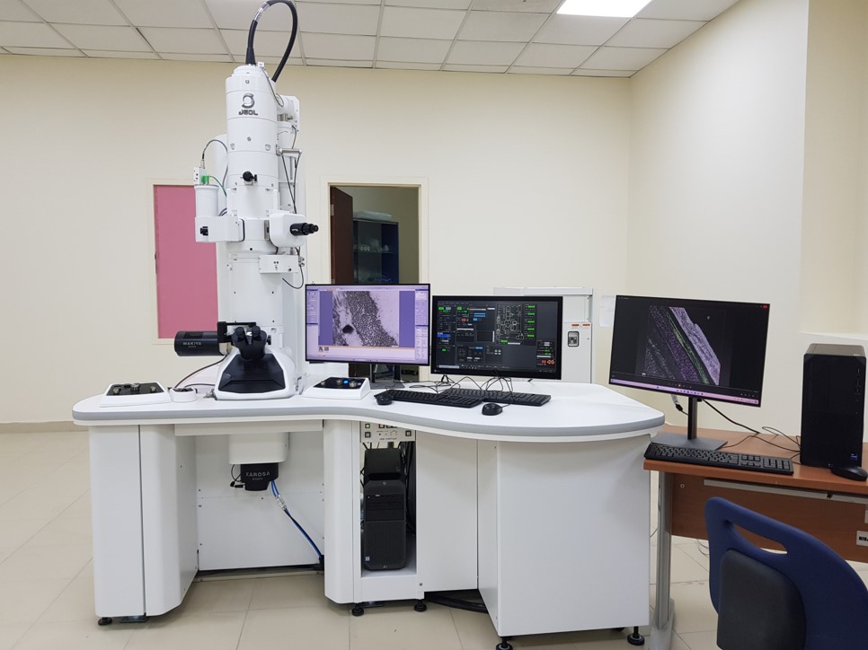
Transmission electron microscope JOEL JEM 1400 (HC)
Xarosa 20 Mega Pixel Bottom Mount CCD Camera
Veleta G3 4 Mega Pixel Side Mount Camera
Specimen Quartet Holder to observe 4 specimens without moving out of the specimen holder
Specimen Quick Change Holder – to observe specimens at a very high magnification (x500,000)
Specimen High Tilting Holder to tilt specimens from -70 degrees to +70 degree to capture 140 images for tomography
Tomography System with computers
Software
Radius software to capture ultrastructural images at a magnification up to a million
Recorder software to capture 140 images at 80,000 magnifications from -70 to +70 degree
Composer software to compose all the 140 images into 3D image format
Visualizer-Kai software to colour code the 3D image
Research
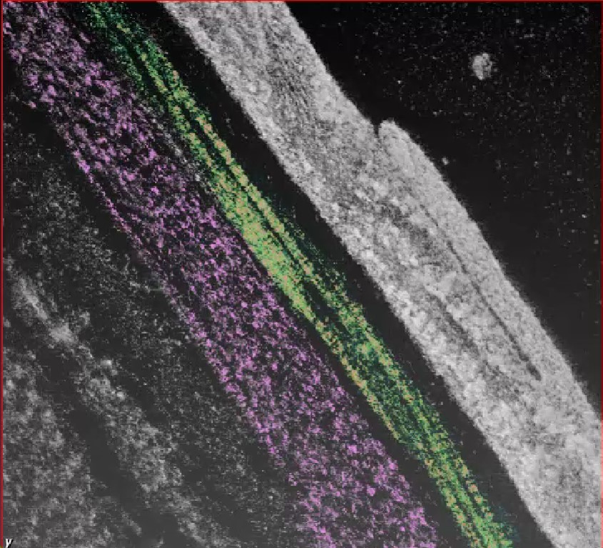
We have expertise to carry out Ultrastructural and 3D tomography research on normal and pathological biological tissue. Our main focus on the ultrastructural organization and 3D electron tomography of collagen fibrils and proteoglycans of normal and pathological cornea such as keratoconus (see picture). We have published our findings in high impact peer-review articles. We also carry out research on the retina, optic nerve, intervertebral disc, knee cartilage and heart valve.
Imaging
3D transmission electron tomography of priendothelial layer (PENL) at of normal cornea
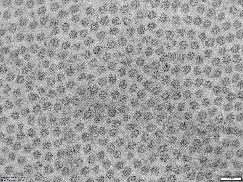
3D transmission electron tomography of collagen fibril of stroma of normal cornea
Uniformly distributed collagen fibril of stroma of normal cornea
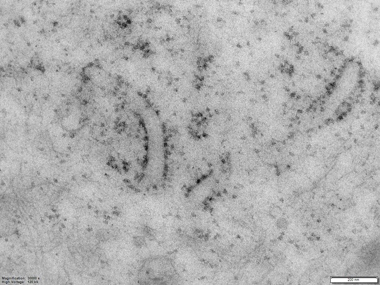
Endoplasmic reticulum of epithelial cell of normal cornea